Continuous Cardiac Output Monitor
June 2023 - Pressent • Stony Brook University • Orthopedic Bioengineering Research Laboratory • Device Design and Prototyping
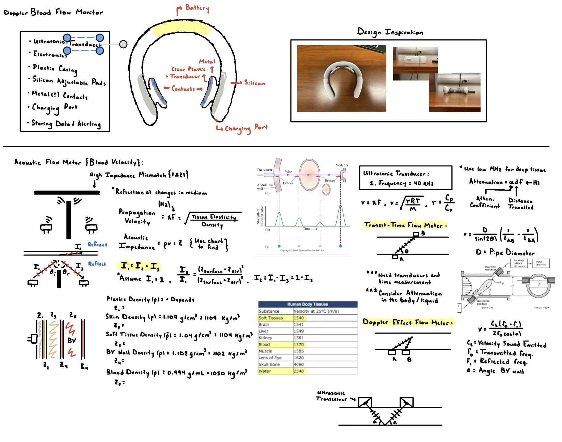
With the Qin Lab at SBU, I am designing an at-home device that continuously monitors cardiovascular health for patient's recovering from intensive conditions (heart failure, heart valve surgery, etc.). As opposed to monitoring the heart's impulse with an EKG, the device relies on ultrasonic technology (in particular develops in flexible ultrasound patches from MIT and UCSD) and arterial tonometry in order to continuously monitor specific benchmarks of cardiovascular health. The project is a part of my undergraduate research as a visiting student of Dr. Qin's lab during the summer semester of 2023 and winter semester of 2024. All the following media is of my own and completed during my time at Stony Brook
Problem Statement and Ideation
This study focuses on developing and testing a feasible prototype design for an at-home, wearable Carotid Artery Cardiac Output Monitor utilizing the Doppler ultrasound and applanation tonometry technology. Severe and sudden myocardial infarction prevails for patients afflicted with recurrent cardiovascular events or recovering from cardiac surgeries, with the mortality rate nearly doubling for patients suffering from recurrent myocardial infarctions after the initial heart attack within a five-year recovery period. Despite the existence of reliable diagnostic technology in emergency-care centers and hospitals, few options offer realistic accessibility of at-home monitoring devices for physiological characteristics of patient’s heart health. However, with recent developments in wearable and flexible ultrasonic transducers from MIT and UC San Diego, the translation of Doppler shift flowmeters into at-home volumetric blood flow monitors is a more realistic opportunity.
Designed to rest on the patient's neck, the device is intended to continuously record data on a patient’s average blood flow through the carotid artery and arterial stiffness through various wearable sensors that are accessible for both doctors and patients through Bluetooth. Together, the Doppler ultrasonic and arterial applanation tonometry technologies provide new methods for developing personalized monitoring of cardiac output for patients diagnosed with chronic cardiovascular disease, recovering from cardiac surgery or an acute cardiovascular event, etc.
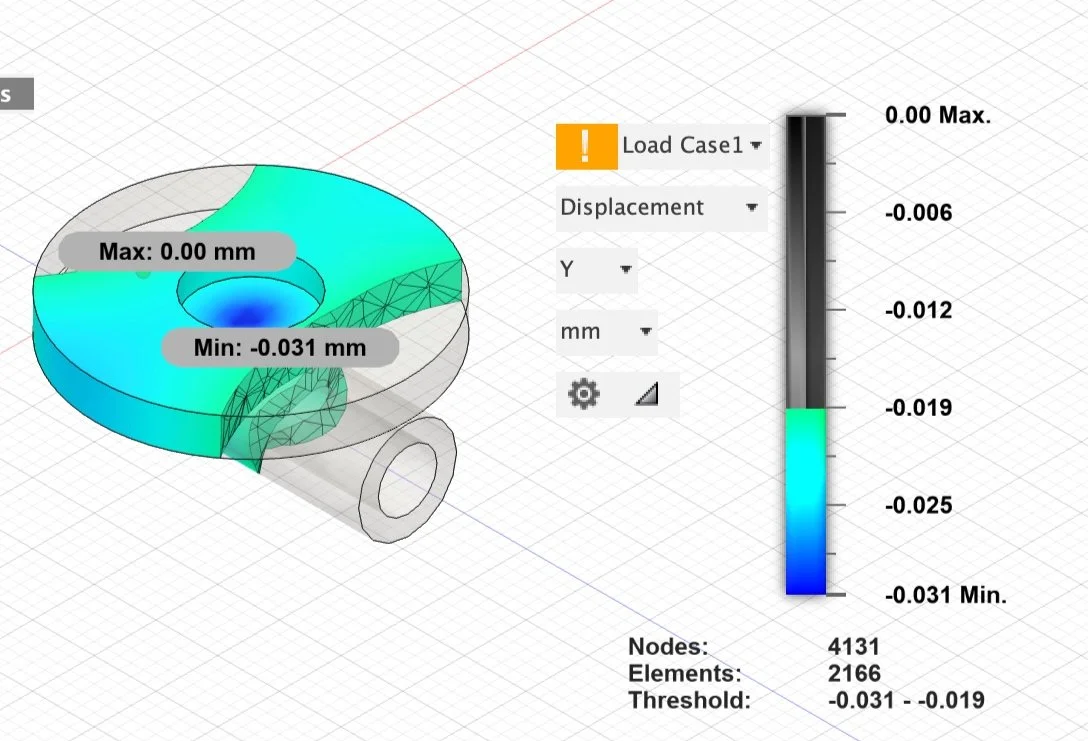
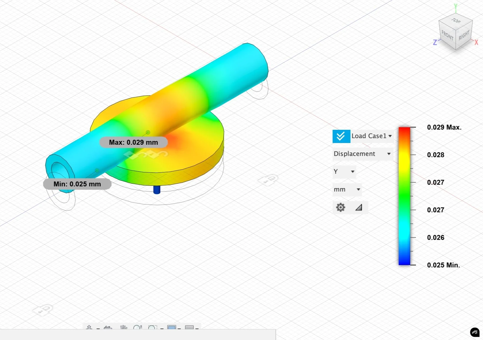
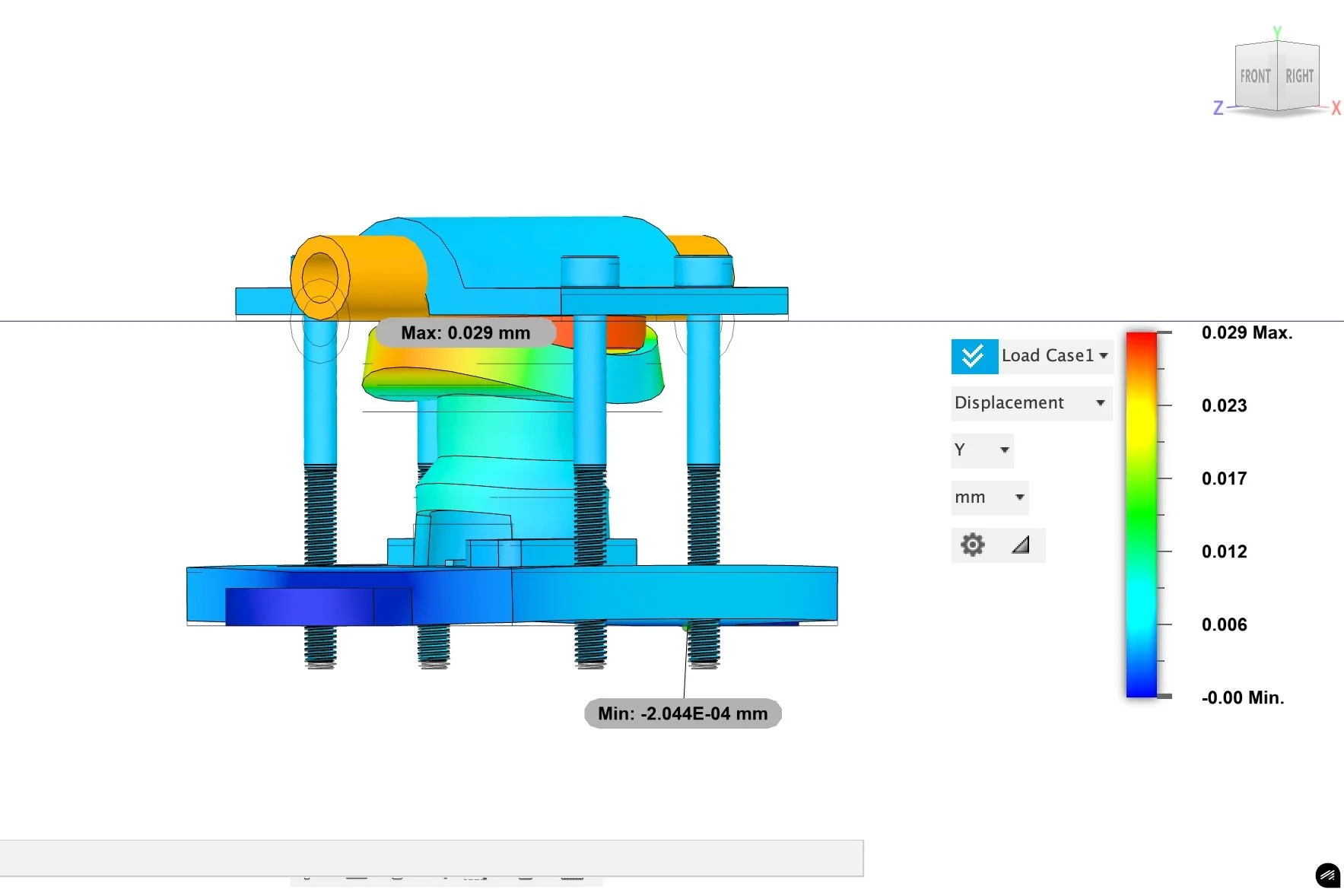
ANSYS Simulation
Ultrasonic Prototyping Process
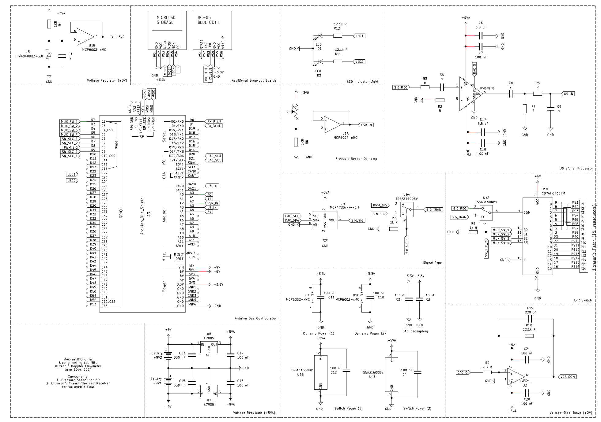
For the ultrasonic patch, the microcontroller controls the signal pulse rate, instantaneous Fast Fourier Transformation (FFT) of the received signal, blood volumetric flow rate calculations, and data storage. A coded sine wave generator on the microcontroller creates a signal with controllable amplitude and frequency that is emitted through the ultrasonic patch. A transmitter and receiver switch switches the patch from transmitter to receiver mode and transmits the received signal to the amplifiers and filters. Finally, a PWM analog pin on the microcontroller receives the signal and executes a program from the “Arduino FFT” library, computing an FFT with the received signal and calculating the instantaneous blood velocity.
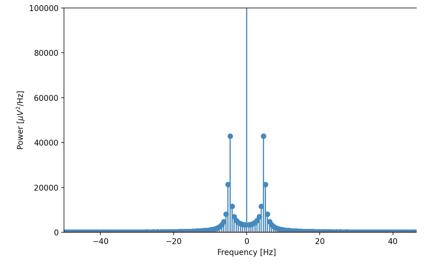
To confirm the validity of the Arduino FFT, a separate Python script was written in order to communicate with the microcontroller’s serial monitor, store the received analog values from the microcontroller, and plot the points. Both a Python Fourier transformation and a hand calculation are conducted to confirm the validation of the transmitted wave.
Prototype Series One
Initial form factors were modeled, indicating the location of the components and the sensors. Based off of a basic neck massager, the design includes suggested materials such as silicones and injection molded ABS plastic, and potential methods for articulating the components.

Prototype Series Three
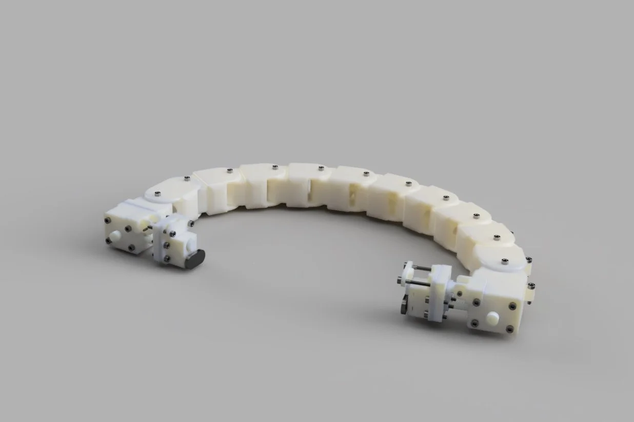
For the final design, this prototype was a tremendous overhaul of the previous two design and incorporates both concerns from the second design stage and greater improvements for manufacturing. Detailed neck links for modular neck adjustment, gear systems for a rack and pinion to engage the sensors, and multiple articulating parks for future adjustments in the design.

Prototype Series Two
Initial prototypes were developed in order to gauge initial concerns with the physical components of the device. The design is the simplest form and includes a living hinge at the center of the back support to provide the means to clamp the device onto the neck. With some cases testing, some notable issues included modularity in the device’s for neck sizes, proper positioning of the sensors relative to the carotid artery, and rigidity.


Blood Pressure Prototyping Process
Arterial Applanation is another recent development in monitoring cardiac output, specifically targeting stiffness and blood pressure. A semi-occlusion method of blood pressure estimation is preferable in the given circumstance considering the continuous application of the device and location of the sensor, and as such arterial applanation is the preferable method.
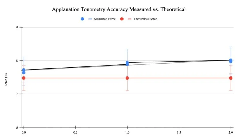
For these probes, a force sensor is utilized, and the resistive change across the sensor is measured and converted with the sensor’s specific resistance-force regression. For the following prototype, testing is conducted with an Ezweiji Film Pressure Sensor with preset resistance-force graphs. The isolated trial apparatus is comprised of a roller pump and reservoir of distilled water, and a vinyl pipe. The figure specifically illustrates the isolated trial for the pressure sensor, which demonstrates the utilization of additional resources such as a specialized pipe clamp to test efficacy.
All components were designed in Fusion360, where ANSYS Static Studies were computed in order to estimate the applanation area with the spring deformation and elastic properties of the vinyl pipe.


